New Patients
(646) 222-6570
Existing Patients
(212) 283-0234

At the office of GD Dentistry, we rely on advanced imaging to guide confident, consistent care. Cone-beam computed tomography (CBCT) gives our team three-dimensional detail that traditional X-rays cannot provide — revealing the shape, position, and relationship of teeth, bone, nerves, and sinuses in a single, highly precise scan. That clarity helps us plan treatments more accurately while minimizing surprises during clinical procedures.
Our practice uses a modern CBCT system selected for image quality, targeted fields of view, and low radiation exposure. The result is fast, reliable imaging that supports a wide range of dental services, from implant placement to the evaluation of complex anatomical issues. We present the results in clear visual formats so patients and clinicians can make informed decisions together.
CBCT transforms a flat X-ray into a complete volumetric picture, allowing clinicians to view cross-sections, axial slices, and 3D reconstructions of the jaws and facial bones. This level of information is especially useful when conventional radiographs leave questions unanswered — for example, about root orientation, bone thickness, or the proximity of anatomical structures that matter for treatment safety.
With these images, clinicians can measure distances and angles with confidence rather than relying on estimation. That precision improves treatment predictability: surgical guides can be planned digitally, endodontic anatomy can be assessed for unusual root configurations, and pathology can be localized with more certainty. In short, CBCT provides the foundation for evidence-based clinical decisions.
Because the images are captured and reviewed digitally, they integrate easily with other diagnostic tools and laboratory workflows. This compatibility facilitates interdisciplinary planning when restorative, periodontal, or surgical specialties must coordinate care, reducing the risk of miscommunication and ensuring everyone is working from the same, accurate dataset.
One of the most common uses for CBCT is implant planning. The scan shows the available bone volume, the quality of that bone, and the relationship to vital structures such as the inferior alveolar nerve and the maxillary sinuses. With this information, the treatment team can choose implant dimensions and positions that maximize long-term stability while avoiding critical anatomy.
CBCT also supports the design of surgical guides that translate digital planning into precise clinical outcomes. Guided implant placement reduces variability, helps maintain proper angulation, and can shorten procedure times by giving the surgeon a clear roadmap. For patients who need complex reconstructions or immediate-loading protocols, the confidence provided by 3D planning is especially valuable.
Beyond implants, CBCT images help evaluate impacted teeth, jaw cysts, and other surgical concerns. When oral surgery is indicated, the ability to visualize the full anatomy beforehand decreases intraoperative guesswork and supports safer, more efficient procedures with predictable healing trajectories.
Patient safety is central to our imaging protocols. Modern CBCT units are designed to limit exposure by using focused fields of view and dose-optimized settings specific to the diagnostic task. Clinicians choose the smallest effective scan volume and the lowest acceptable exposure that will still provide diagnostically useful information, in keeping with the ALARA (as low as reasonably achievable) principle.
Proper training and protocol selection are as important as the equipment itself. Our team evaluates each case individually to determine whether a CBCT scan is necessary and which parameters will best answer the clinical question. This selective approach ensures that patients receive the diagnostic benefits of three-dimensional imaging only when it will influence treatment decisions or improve outcomes.
The scans are interpreted with attention to both dental and maxillofacial anatomy; any unexpected findings outside the immediate dental concern are documented and communicated. If additional evaluation by a specialist is needed, we coordinate next steps to ensure comprehensive follow-up while keeping patient welfare a priority.
CBCT’s value extends well beyond surgical planning. In endodontics, it reveals complex root canal morphology, locates hidden canals, and helps detect periapical lesions that may be obscured on standard films. For orthodontic cases, three-dimensional images assist in assessing jaw relationships, impacted teeth, and airway space to inform safer and more effective movement strategies.
Airway assessment is another growing application. While CBCT is not a standalone diagnostic tool for sleep apnea, it provides structural information about the nasal passages, pharyngeal airway, and skeletal contributors that can inform referral decisions or adjunctive treatment planning. Similarly, periodontal clinicians can evaluate bone defects and furcation involvement with greater accuracy when 3D data are available.
Because the scan captures the surrounding soft-tissue outlines and bone architecture, it also aids in identifying less common conditions such as cysts, benign tumors, or developmental anomalies. Detecting these issues early supports timely referral and treatment when necessary, illustrating how CBCT contributes to comprehensive oral health care.
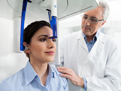
A CBCT scan is quick and noninvasive. On arrival, a member of the team will review your medical history and explain the reason for the scan so that the correct field of view and exposure settings are chosen. You will be seated or positioned standing, depending on the unit, and asked to remain still for the brief acquisition — typically under a minute for most targeted scans.
The process requires no special preparation in most cases; removable metal objects such as glasses or jewelry are taken off to prevent artifacts in the image. Patients who have mobility considerations receive individualized accommodations to ensure comfort and accurate positioning. After the scan, the images are processed and reviewed by the clinician, who will explain the findings and how they relate to your treatment options.
Because CBCT captures detailed volumetric data, it is often possible to share clear visual explanations with patients, including cross-sectional views and 3D reconstructions. This visual approach helps clarify complex issues and supports shared decision-making, so you can participate fully in selecting the path that best meets your health and functional goals.
In summary, cone-beam computed tomography is a powerful diagnostic tool that enhances clinical accuracy, promotes safety through targeted imaging, and supports better-informed treatment planning across many dental specialties. If you would like to learn more about how CBCT may be used in your care, please contact GD Dentistry for additional information.
Cone-beam computed tomography, or CBCT, is a three-dimensional imaging modality that captures volumetric views of the teeth, jaws, and surrounding facial structures. Unlike traditional two-dimensional dental X-rays, CBCT acquires a full 3D data set that can be reconstructed into axial, sagittal and coronal views for detailed assessment. These 3D images reduce overlap and distortion, making it easier to visualize anatomic relationships and complex structures.
At GD Dentistry we use CBCT to complement standard radiography when additional diagnostic detail is needed for treatment planning. CBCT is not a replacement for routine 2D X-rays in every visit, but it is the preferred tool when precise spatial information is required for diagnosis or surgery.
CBCT provides the detailed anatomic information necessary to plan implant placement with high precision, including bone volume, bone density, and the location of critical structures such as nerves and the sinus cavities. These 3D data sets allow clinicians to measure dimensions precisely and to select implant size and angulation that match the patient’s anatomy. Accurate preoperative planning reduces the risk of complications and supports predictable restorative outcomes.
Surgeons can also use CBCT images to create surgical guides and to simulate implant positioning before the procedure. This level of preparation improves efficiency during surgery and helps ensure the implant integrates properly with the available bone.
CBCT uses a focused cone-shaped X-ray beam and produces a lower radiation dose than conventional medical CT scans while delivering far more diagnostic information than standard dental X-rays. Modern CBCT units and optimized scanning protocols further minimize exposure by limiting the field of view to the region of interest and using the lowest effective dose for diagnostic needs. As with any imaging, the decision to use CBCT is based on clinical benefit versus exposure, and imaging is only recommended when it will influence diagnosis or treatment.
Your dentist will select appropriate scan settings and shielding when necessary to follow the principle of ALARA (as low as reasonably achievable). If you are pregnant or have specific medical concerns, inform the dental team so they can take extra precautions or consider alternative diagnostic options.
A CBCT appointment is typically quick and noninvasive. The patient is positioned in the machine—either sitting, standing, or in some models lying down—and asked to remain still for the duration of the scan, which generally takes less than a minute of exposure time and a few minutes for overall setup and positioning. There is no mouthpiece or intraoral sensor required for the volumetric scan, and most patients experience no discomfort during the procedure.
After the scan, the images are processed and reviewed by the dentist, often during the same visit, so they can discuss findings and next steps with you. Because the data are three-dimensional, the clinical team can manipulate the images to examine specific locations from multiple angles and plan treatment precisely.
CBCT is a versatile diagnostic tool that aids in evaluating a wide range of conditions including impacted or malformed teeth, root fractures, complex endodontic anatomy, temporomandibular joint disorders, sinus pathology, and bony lesions. The ability to visualize anatomy in three dimensions allows clinicians to detect issues that can be missed or obscured on traditional 2D images. For example, small root fractures, accessory canals, or the proximity of pathology to anatomical landmarks can be assessed more reliably with CBCT.
These detailed images support more accurate diagnoses and targeted treatment planning across dental specialties, from endodontics to oral surgery and orthodontics. When additional consultation is needed, CBCT images can be shared with specialists to coordinate comprehensive care.
Yes, CBCT is often used to assist in orthodontic treatment planning and in the evaluation of airway anatomy. The 3D data allow clinicians to assess tooth positions, root alignment, jaw relationships, and facial asymmetries with greater clarity than 2D records. For airway assessment, CBCT can reveal constrictions, relative airway volume, and anatomical contributors to obstructive sleep apnea that may inform multidisciplinary treatment recommendations.
CBCT findings are interpreted in the context of a full clinical examination and other diagnostic records. Orthodontists and sleep specialists may combine CBCT data with clinical assessments and functional testing to develop a comprehensive treatment plan tailored to each patient’s needs.
Image acquisition itself is rapid, and processing typically completes within minutes, allowing the dental team to begin reviewing the volumetric data shortly after the scan. A thorough interpretation may take longer depending on the complexity of the case and whether multiplanar reconstructions, cross-sections, or 3D renderings are required. The dentist will analyze the images to identify anatomy, pathology, and measurements critical to the proposed treatment.
Once interpreted, CBCT images are integrated into the procedural workflow, whether that means designing a surgical guide for implant placement, planning a root canal approach, or coordinating care with a specialist. Patients are then informed of the findings and the recommended treatment steps based on objective imaging evidence.
CBCT should be used selectively and only when the expected diagnostic benefit outweighs the radiation exposure. For routine examinations or simple problems that can be adequately evaluated with conventional 2D radiography, CBCT is generally not indicated. Special consideration is given to children, pregnant patients, and patients with unique medical conditions; in such cases, dentists will weigh alternatives and adjust protocols to minimize exposure.
Your dental team will review your medical history and current health status before recommending CBCT, and they will explain why an advanced 3D scan is or is not necessary for your situation. If a different imaging approach is more appropriate, the clinician will suggest the safest effective option.
Accurate interpretation of CBCT images depends on proper image acquisition, appropriate field of view selection, and clinician expertise in reading volumetric data. Dentists who use CBCT are trained to recognize normal anatomical variations, artifacts, and the limitations of the modality, and they apply systematic review protocols to avoid missed findings. When needed, clinicians may consult with or refer images to specialists such as oral and maxillofacial radiologists for advanced interpretation.
Quality assurance also involves regular maintenance and calibration of the CBCT unit, adherence to recommended scanning parameters, and the use of validated software tools for measurements and surgical planning. These practices help ensure that CBCT contributes reliable information to clinical decision-making.
Yes, CBCT images are often shared with other dental or medical specialists to support coordinated care when a referral is necessary. Digital volumetric files and accompanying reports can be exported and transmitted securely to collaborating clinicians so they can review the same detailed information when planning treatment. Sharing images improves communication and helps ensure all providers are working from a consistent diagnostic data set.
Your dental team will request your consent before sending images to outside providers and will take steps to protect your privacy in accordance with applicable regulations. If you prefer, copies of your CBCT data can also be provided to you for transfer to another clinician.