New Patients
(646) 222-6570
Existing Patients
(212) 283-0234

Digital radiography replaces traditional film with electronic sensors and computer processing to capture dental X-ray images. Instead of developing film in a darkroom, the sensor records the image and sends it instantly to a computer where clinicians can view, enhance, and store the result. This shift from chemical film to digital files has changed how dentists gather diagnostic information, streamlining workflows while preserving the level of detail clinicians need to evaluate teeth, bone, restorations, and other oral structures.
For patients, the practical benefits are immediately noticeable: images appear on-screen within seconds, adjustments such as contrast and magnification can be made without retaking images, and files are easily archived for future comparison. These capabilities help clinicians spot subtle changes over time and make informed decisions sooner. In a modern dental practice, digital radiography is not just a convenience — it is a foundational tool that supports accurate diagnosis and efficient treatment planning.
Beyond the technology itself, the broader value lies in improved communication. High-resolution images help clinicians explain conditions and proposed treatments more clearly to patients, and electronic files simplify secure sharing with specialists when collaborative care is needed. When used thoughtfully, digital radiography enhances both the clinical process and the patient experience.
Contemporary digital sensors come in a range of sizes and designs that are less intrusive than older film packets. Many sensors are thinner and more flexible, which reduces gag reflex triggers and makes intraoral positioning easier for both front and back teeth. Because sensors provide immediate feedback, clinicians can confirm image quality on the spot and correct positioning without additional exposure delays, reducing the need for repeat images.
Another advantage is the ability to fine-tune images through software. Clinicians can adjust brightness, contrast, and sharpness, or apply measurement tools to assess bone levels and root lengths. These enhancements do not change the underlying anatomy but make diagnostically relevant features easier to interpret. In practice, this means more reliable detection of cavities, fractures, and periodontal issues at earlier stages.
Patient comfort and precision often work together: better-fitting sensors and quicker capture mean shorter appointments and a smoother imaging experience. For patients with anxiety or physical limitations, the reduced time and improved ergonomics of digital systems can make routine X-rays a more tolerable part of care.
Instant image availability shortens the gap between capture and diagnosis. Rather than waiting for film to be processed, clinicians view digital images immediately and can discuss findings with the patient right away. This accelerates decision-making and allows clinicians to combine image review with intraoral examinations and other evaluations during the same visit, creating a more cohesive appointment flow.
Digital files also facilitate efficient collaboration. When a case requires input from a specialist or a laboratory, electronic images can be shared securely and quickly, enabling interdisciplinary planning without postal delays or the need to transport physical film. This smoother exchange helps ensure that referrals contain precise information and that treatment steps are coordinated across providers.
Electronic storage makes longitudinal comparison straightforward: prior images are readily available to track healing, assess the stability of restorations, or monitor progressive conditions. That continuity supports preventive care and helps clinicians tailor interventions based on clear visual trends rather than isolated snapshots.
One of the most important benefits of digital radiography is reduced radiation exposure compared with traditional film techniques. Digital sensors are typically more sensitive to X-rays, so diagnostic-quality images can be obtained using lower doses of radiation. Clinicians still follow the principle of ALARA (as low as reasonably achievable), but digital systems provide an extra margin of safety that is especially valuable for patients who need periodic imaging.
Digital imaging also eliminates the need for chemical processing, which has both practical and environmental implications. Film development requires developer and fixer solutions that must be handled and disposed of according to regulatory guidelines. By removing that step, digital workflows reduce chemical waste and the logistical burden of managing hazardous byproducts, aligning dental operations with broader sustainability goals.
Safety procedures remain a priority: lead aprons, thyroid collars when appropriate, and strict exposure protocols continue to be standard practice. Digital radiography augments these safeguards by combining sensitive sensors with faster capture and fewer repeat exposures, contributing to a safer diagnostic environment overall.
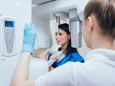
During a typical digital X-ray visit, a member of the clinical team will explain the process and position a sensor in the mouth or place a small external device for extraoral views, depending on the type of image needed. The sensor is secured briefly while the image is taken, and the exposure itself lasts only a fraction of a second. Because the image appears on screen immediately, the clinician can confirm quality and proceed without delay.
Clinicians use the on-screen image to point out relevant findings and to answer patient questions in real time. If further views are required, adjustments can be made quickly and efficiently. For patients who feel anxious, the brief exposure time and fewer repeats make the experience less taxing than older film-based methods, while still delivering the diagnostic detail clinicians rely on.
After capture, images are saved to the patient’s electronic record and retained for future comparisons. If specialist input is needed, the office can securely share the images to support collaborative care. Overall, the process is designed to be fast, transparent, and focused on gathering the information necessary to recommend safe, effective treatment.
At the office of GD Dentistry, digital radiography is one of several technologies we use to support precise, patient-centered care while minimizing inconvenience and exposure. If you have questions about imaging or want to learn how digital X-rays fit into your treatment plan, please contact us for more information.
Digital radiography uses electronic sensors and computer technology to capture, display, store and share dental x-ray images. Instead of traditional film, a sensor or plate records the image and transmits it instantly to a computer for review. This workflow shortens appointment time and allows clinicians to enhance images for clearer viewing.
Captured images are saved directly into the patient record so they can be retrieved, compared and printed as needed. Because no film or chemical processing is required, digital radiography is also more environmentally friendly than conventional film systems. The immediate availability of images supports faster diagnosis and treatment planning.
Digital sensors are more sensitive to x-rays than traditional film, so they require a lower dose to produce a diagnostically useful image. Advanced software can further enhance detail and contrast, which reduces the need for repeat exposures. The ability to review images immediately also helps the clinician confirm correct positioning on the first attempt.
Clinics follow the ALARA (as low as reasonably achievable) principle to minimize exposure, using collimation, appropriate exposure settings and protective shielding. Special techniques and smaller sensors for pediatric patients further limit dose when imaging children. Overall, digital systems allow clinicians to obtain the necessary diagnostic information with less radiation than film-based methods.
Digital dental x-rays are taken with an intraoral sensor or an extraoral digital detector positioned near the teeth or head, depending on the type of image required. The sensor captures the x-ray image and transmits it directly to imaging software on a computer, where the clinician can view it immediately. Proper positioning and bite registration are used to ensure diagnostic images with minimal retakes.
Once the image appears on screen, the clinician can adjust brightness, contrast and magnification to evaluate structures more precisely. Images are labeled and attached to the patient file in the practice management system for long-term record keeping. Many systems save images in standard formats that can integrate with other imaging modalities and treatment planning software.
Digital x-rays reduce radiation exposure compared with traditional film, which makes them a safer option for both children and adults when imaging is clinically indicated. For pediatric patients, smaller sensors and child-specific exposure settings are used to limit dose while still obtaining necessary diagnostic information. Protective measures such as lead aprons and thyroid collars are applied whenever appropriate.
For patients who are pregnant or may be pregnant, dentists generally avoid routine radiographs unless the information gained will affect urgent care, and they take extra precautions when imaging is necessary. It is important to inform the dental team about pregnancy so they can adjust protocols and use shielding to protect the abdomen and thyroid. Decisions about imaging are always made with patient safety and clinical need in mind.
Digital radiographs provide clear two-dimensional views of tooth roots, surrounding bone and periodontal structures that are essential for preliminary implant evaluation and restorative planning. When combined with three-dimensional imaging such as CBCT, clinicians gain more complete information about bone height, width and anatomical landmarks needed for implant placement and full mouth reconstruction. Digital images facilitate precise measurements and preoperative assessment.
These images can be used alongside digital treatment-planning tools to map implant positions and design provisional restorations, improving coordination between the clinician and the dental laboratory. The digital workflow reduces manual steps and supports more predictable communication among the treatment team. Images also serve as baseline records to monitor healing and long-term outcomes.
Digital radiographs are archived electronically within the practice management and imaging systems, where they are linked to each patient record for organized retrieval. Clinics implement routine backups and use secure servers or cloud services to prevent accidental data loss. Electronic storage eliminates the need for physical film libraries while improving access to historical images.
Access controls, user authentication and encryption are common measures used to protect patient data and comply with privacy regulations. Practices keep audit logs to track who accessed or modified records and use secure transfer methods when sharing images with outside providers. At GD Dentistry, standard procedures are followed to safeguard imaging data and patient health information.
Digital x-rays can be displayed instantly on a chairside monitor, allowing clinicians to review images with patients during the same visit. Image enhancement tools such as zoom, brightness and contrast adjustments help reveal subtle findings that might be harder to see on film. This immediacy and clarity support faster, more accurate diagnosis.
Being able to point out specific areas on the screen helps patients understand their condition and the rationale for recommended treatment. Digital files also allow practitioners to annotate images, generate visual treatment plans and compare images over time to document disease progression or healing. At GD Dentistry, clinicians use these capabilities to involve patients in informed discussions about their care.
Digital radiography eliminates the need for chemical developers and fixer solutions that are required for processing film, reducing the generation of hazardous waste. It also removes the need for physical film and paper records, which decreases material consumption and storage demands. These changes contribute to a smaller environmental footprint for dental imaging operations.
Electronic transfer of images reduces the need for shipping physical films between offices or to specialist labs, further lowering resource use and carbon emissions. Many practices adopt digital workflows as part of broader sustainability initiatives to reduce waste and improve operational efficiency. The absence of chemical processing also simplifies compliance with environmental disposal regulations.
Yes, digital images can be shared electronically with specialists, laboratories or referring dentists using secure transfer protocols and standardized file formats. Common formats such as DICOM allow images to be imported into different imaging and planning systems, preserving diagnostic information and metadata. Electronic sharing speeds up consultations and coordination of care compared with mailing physical films.
When sending images outside the practice, clinics typically use encrypted portals, secure email or direct system integrations to protect patient privacy. Patient consent and documentation are part of the process to ensure compliant information exchange. Timely sharing of images helps specialists provide informed recommendations and supports multidisciplinary treatment planning.
Patients should bring any recent dental x-rays or imaging records if they are available, as prior images can help the clinician compare changes and avoid unnecessary repeats. Wear comfortable clothing and avoid jewelry or removable oral appliances that could interfere with imaging. Inform the dental team about pregnancy, recent medical changes or specific concerns that may affect imaging choices.
Expect the imaging process to be quick; sensors are positioned and exposures are taken in moments, with images available immediately for review. If specific images are planned, the staff will explain steps and protective measures such as lead aprons and thyroid collars. Patients are encouraged to ask questions about what the images show and how they will be used in diagnosis and treatment planning.